MAKE A MEME
View Large Image

| View Original: | Endometriosis, abdominal wall 2.jpg (650x432) | |||
| Download: | Original | Medium | Small | Thumb |
| Courtesy of: | commons.wikimedia.org | More Like This | ||
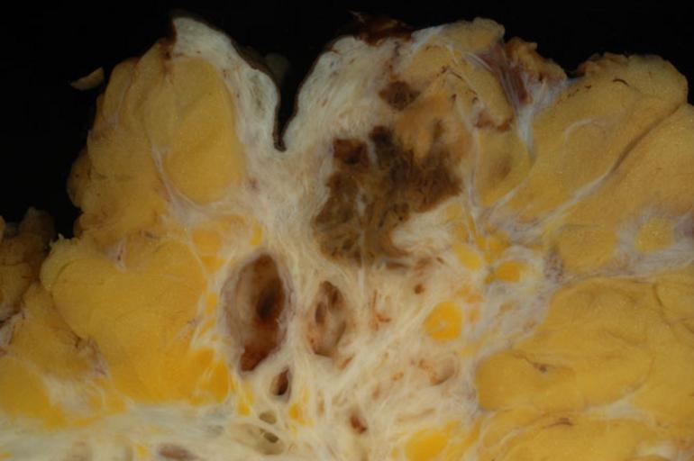
| Keywords: Endometriosis, abdominal wall 2.jpg The photo shows the same case presented in Image Endometriosis abdominal wall jpg For the photo I moved in a little closer and immersed the specimen in a shallow pan of water Whether it's better to shoot specimens dry or immersed is a matter of taste I prefer the more natural look of immersed specimens but others like the sharpness of dry surfaces If you do shoot specimens dry be sure to sop as much moisture off the surface as possible Otherwise distracting highlights will ruin the picture Both photos were taken with a Nikon D100 digital SLR camera through a Sigma 50mm macro lens The camera was set for manual focus incandescent light balance and aperture priority exposure mode at f/8 Both shots looked a little dark on the computer monitor so I expanded the dynamic range in Photoshop using the Levels command The only other digital editing was slight cropping of the top photo and downsampling to proper size 650 pixels greatest linear dimension for screen display There was no sharpening or color correction The imaged was saved in JPEG format with the quality setting 8 High The black background is a piece of velvet Photograph by Ed Uthman MD Public domain Posted 22 May 04 http //web2 airmail net/uthman/specimens/index html PD Ed Uthman Gross pathology Endometriosis | ||||