MAKE A MEME
View Large Image

| View Original: | Intravascular Leiomyomatosis of the Uterus.jpg (650x432) | |||
| Download: | Original | Medium | Small | Thumb |
| Courtesy of: | commons.wikimedia.org | More Like This | ||
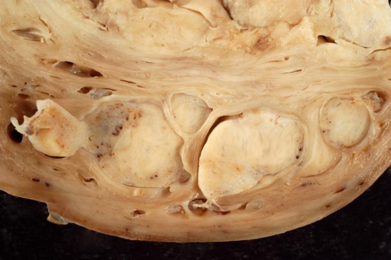
| Keywords: Intravascular Leiomyomatosis of the Uterus.jpg This patient underwent hysterectomy for fibroids However when the gynecologist exposed the uterus there were what appeared to be distended blood vessels extending from the lower uterine segment into the parametria It was not technically feasible to resect the entire uterus so a supracervical hysterectomy was performed On gross exam the fundus was dominated by a 10-cm leiomyoma However most remarkable was the presence of numerous markedly distended blood vessels in the myometrium containing firm rubbery tumors that were otherwise identical to the main tumor In the photo above the main tumor intrudes into the frame from above while several markedly dilated veins distended with tumor are seen in the bottom half This photo was taken with a Nikon D-100 digital SLR fitted with a Sigma 50-mm macro lens set at f/8 The image was captured in RAW mode but I later downsampled it considerably in the interest of minimizing file size Photograph by Ed Uthman MD Public domain Posted 17 Jul 2005 http //web2 airmail net/uthman/specimens/index html PD Ed Uthman Gross pathology of neoplasms Uterine fibroids | ||||