Click Here for More Images from iStock- 15% off with coupon 15FREEIMAGES 
\
Young female scientist working at the laboratory with an electron microscope
A close-up hand of researcher inserting a microscope slide into a microscope within a laboratory. Precision and care required in scientific research, emphasizing the critical role of laboratory equipment in the analysis and discovery processes
close-up microscope isolated on black background
CNC surface grinding machine industrial exhibition
Modern microscope equipped with a digital camera, computer and monitor in a biological research laboratory. Modern microscope with digital imaging system
Closeup of Scientific microscope data analysis in the laboratory, medicine equipment research setting in lab for chemistry analysis
Scanning Electron Microscope (SEM) machine in laboratory and blur computer controller background.
Optometrist conducting automated refraction test. Modern eye examination equipment in use for vision diagnosis.
3D laser microscope measuring PCB board. Industrial manufacturing
Microscope in medicine, black and white image
Latin American optometrist performing an eye exam on a patient using an eye scanner
Testing of electronic equipment
Disassembled electronic circuit assembly for amp repair black and white
Circuit board is tested on the device to identify assembly problems
This is a clean and disassemble the lens for photography into its component parts
Chemical laboratory technician holding a microscope slide in laboratory.
Profile projector dimension measurement machine measuring workpiece dimension in second. Industrial manufacturing
Connection joint of stainless steel pipe in fectory
Ophthalmology machine equipment for testing vision with blurred blue light background
cross section show metal helical gear or cogs gear bearing housing shaft and detail inside of transmissions gearbox for industrial assembly with electric induction motor
Medicine, Plasma, Equipment, Liquid, Pharmacy
Vintage Stereo Turntable Vinyl Record Tonearm Mechanism Closeup. Shiny Metal Parts of the Complex Mechanical Component.
Laboratory equipment in the hospital laboratory department
Pharmaceutical scientist working in an aspeptic or sterile or clean room
old photo camera with bellows and lens in detail
CAD CAM dental computer-aided machine. Digital modern dental laboratory for prosthesis and crowns milling
Close-up on a microscope at the laboratory - science concepts
Scientist is preparation of nanomaterials for Scanning Electron Microscope (SEM) machine in laboratory
Close-up of optometry slit lamp in a doctors office
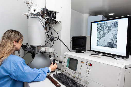
Free Images: "bestof:Scanning Electron Microscope with Spin Polarization Analysis (5941086294).jpg This NIST-developed instrument a scanning electron microscope with spin"
Leaf epidermis.jpg
Butterfly tongue.jpg
cells-electron-microscope-macro-570676.jpg
bacteria-electron-microscope-811861.jpg
bacteria-electron-microscope-808154.jpg
bacteria-electron-microscope-stained-808158.jpg
pollen-microscope-543490.jpg
hela-cells-cultured-544312.jpg
salmonella-bacteria-macro-549608.jpg
citrus-yellow-mite-macro-989500.jpg
cancer-cells-cells-scan-541954.jpg
pestis-bacteria-bubonic-plague-541948.jpg
blood-cells-cells-human-543484.jpg
hela-cells-bacteria-germs-virus-544318.jpg
bacteria-disease-escherichia-coli-62993.jpg
mite-aceria-anthocoptes-acari-67638.jpg
yellow-mite-macro-lorryia-formosa-847862.jpg
white-blood-cell-cell-blood-cell-543471.jpg
koli-bacteria-escherichia-coli-123081.jpg
yellow-mite-macro-lorryia-formosa-847873.jpg
microscope-lab-research-medical-316556.jpg
Ebola_Virus_Particles.jpg
Misc pollen colorized.jpg
Ebola_Virus_Particles.jpg
Misc pollen.jpg
Signale REM.svg
Pear interaction SEM german.svg
Laughlin quasiparticle interferometer sample scanning electron micrograph.svg
tin-spheres-colorized-1444073.jpg
Pear interaction SEM english.svg
Electrically detected magnetic resonance.svg
Fine hyperfine levels.svg
Andreev reflection.svg
Constant current.svg
Constant height.svg
Elaine_Zworykin.jpg
Spinning_Reels_Maze.jpg
Hepatitis B virus v2.svg
Surface ionization of cesium.svg
P450cycle.svg
Scanning Electron Microscope with Spin Polarization Analysis (5941086294).jpg
Leaf epidermis w scale.jpg
Walnut leaf 23 002.jpg
Walnut leaf 23 001.jpg
രക്തകോശങ്ങൾ.jpg
Leukocyt.png
Chemical Analysis;Maximum Pixel Software (5884305705).jpg
Chemical Analysis; Magnesium Oxide Dice (5887269391).jpg
Red_White_Blood_cells.png
Red White Blood cells.jpg
Scanning Electron Microscope Kemira.jpg
Curculionidae.jpg
Chlamydomonas6-1.jpg
Chlamydomonas2-4.jpg
Chlamydomonas2-1.jpg
Chlamydomonas2-2.jpg
Chlamydomonas2-3.jpg
Chlamydomonas2-5.jpg
Chlamydomonas2-6.jpg
Chlamydomonas2-7.jpg
Chlamydomonas2-8.jpg
HederelloidSEM.jpg
Lampyridae.jpg
Microblinds.JPG
Microblind.JPG
Actinedida.jpg
Aphaenogaster.tif
Velvet_ant_stinger_(tip)--scanning_electron_microscope_image.jpg
Blood clot in scanning electron microscopy.jpg
Aphid micro.jpg
Tingidae family.jpg
Miridae SEM 2.jpg
Miridae SEM 3.jpg
Lodewyck2.jpg
SEM school logo.jpg
Micro-blinds.JPG
Carpenter Ant.jpg
Bronchiolar epithelium 1 - SEM.jpg
Bronchiolar epithelium 2 - SEM.jpg
Bronchiolar epithelium 3 - SEM.jpg
Bronchiolar epithelium 4 - SEM.jpg
Oxytricha_trifallax.jpg
Clay_magnified.jpg
Nanowires (5887263715).jpg
Rayon_flock_ESM.png
SEM_flock_particles.png
Nanotube Soot (6648549965).jpg
Nanowire; Sensor (5978119246).jpg
Remf dartmouth edu curculionidae.jpg
Agaricus bisporus spores SEM 1.jpg
Agaricus bisporus spores SEM 2.jpg
Agaricus bisporus spores SEM 3.jpg
Coleus leaf trichomes SEM.jpg
Carbon Nanotube Measurements (5887265427).jpg
Conductive probe SEM images.png
Spermatozoa-human-3140x.jpg
Spermatozoa-human-1000x.jpg
Nanoscale Line Roughness (5884303799).jpg
Silicon Rings (10747924673).jpg
Drosophilidae compound eye.jpg
Terms of Use   Search of the Day